The right scan depends on the question
There is no single best heart scan. An echocardiogram, cardiac CT and cardiac MRI each answer different questions. The most useful test depends on what your doctor is trying to find out: valve function, heart muscle strength, coronary artery disease, scarring, inflammation, or structural anatomy.
This is why imaging should start with the clinical problem, not with the machine. A patient with exertional chest tightness may need assessment for coronary artery disease. A patient with a murmur may need a detailed valve assessment. A patient with unexplained breathlessness may need an echocardiogram first, and sometimes MRI afterwards.
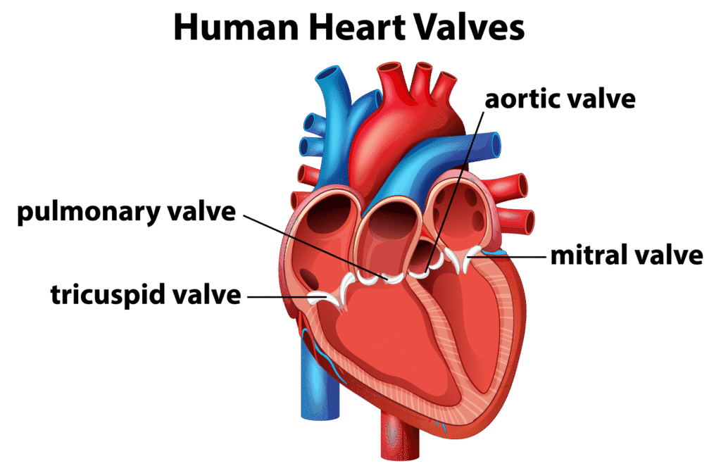
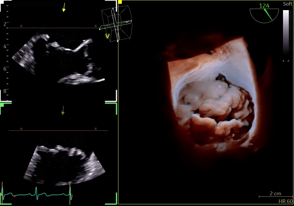
Echocardiogram: heart ultrasound
An echocardiogram uses ultrasound to show the heart moving in real time. It is usually the first-line imaging test for many cardiology symptoms because it is quick, safe and gives immediate information about the heart’s structure and function.
It is especially useful for assessing:
- heart valve narrowing or leakage, including heart valve disease;
- heart pumping strength, including ejection fraction;
- heart chamber size;
- fluid around the heart;
- some congenital abnormalities;
- causes of breathlessness or suspected heart failure.
The main limitation is that ultrasound does not directly show the coronary arteries. Image quality can also vary depending on body shape, lung disease and the acoustic windows available.
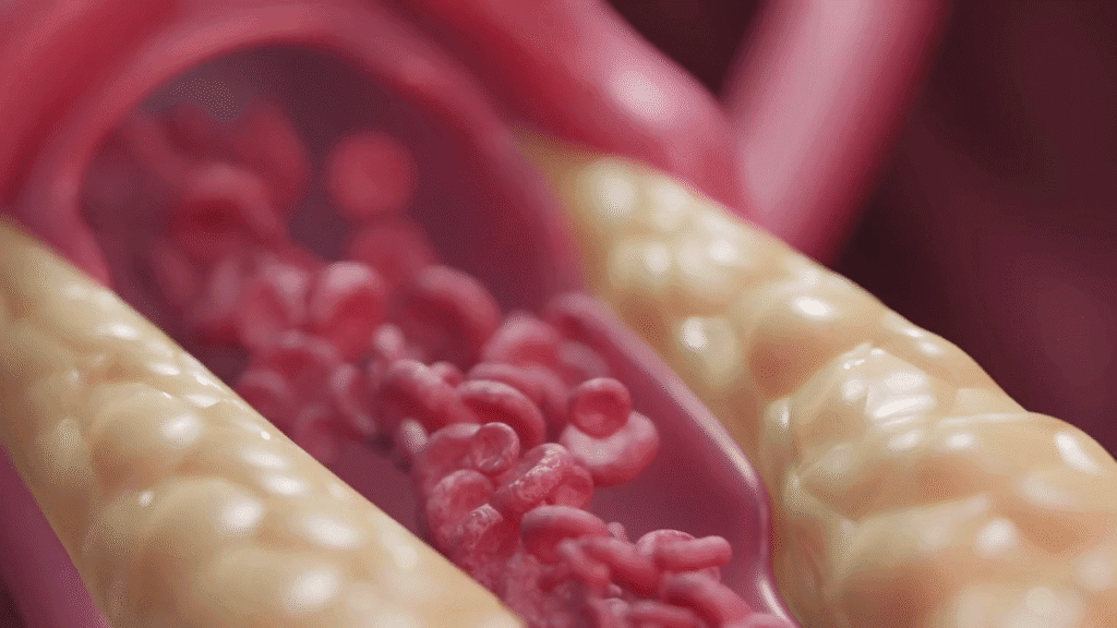
Cardiac CT: coronary arteries and anatomy
Cardiac CT uses X-rays to build detailed images of the heart and coronary arteries. It is often used when the question is whether cholesterol plaque or narrowing is present in the coronary arteries.
It is useful for:
- investigating possible angina or coronary artery disease;
- calcium scoring and cardiovascular risk assessment in selected patients;
- planning some valve procedures;
- mapping complex cardiac anatomy.
Cardiac CT involves a small dose of radiation and often uses contrast dye. It may not be suitable for everyone, particularly patients with severe kidney impairment or an irregular/fast heart rhythm that prevents clear images.
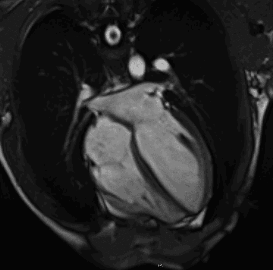
Cardiac MRI: heart muscle detail
Cardiac MRI uses magnets and radio waves rather than radiation. It is particularly useful when the question concerns the heart muscle itself.
MRI can help assess:
- heart muscle scarring;
- myocarditis or inflammation;
- cardiomyopathy;
- viability after reduced blood supply;
- heart volumes and function with high precision;
- complex congenital or structural problems.
It takes longer than echo or CT and is not suitable for all patients with certain metal implants or severe claustrophobia.
How the tests fit together
In real practice, these scans often complement each other. Echo may identify valve disease. CT may define anatomy before a procedure. MRI may show whether the heart muscle has started to scar or remodel. The best imaging strategy is the one that answers the clinical question with the least unnecessary testing.
Dr Mark Cassar is accredited in echocardiography, cardiac CT and cardiac MRI. That means he can advise on which test is most appropriate and interpret results in clinical context rather than treating each scan as a separate event.
When to discuss heart imaging
If you have chest pain, breathlessness, palpitations, a murmur, abnormal ECG findings or an unclear previous scan result, a cardiology review can help decide which test is needed. Details of imaging available through Dr Cassar’s private practice are on the cardiology services page, with appointment information available here.