What is a cardiac MRI?
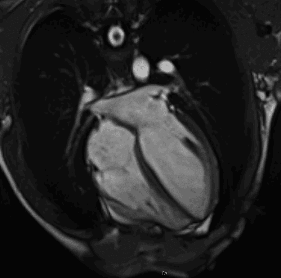
A cardiac MRI is a detailed scan of the heart muscle, valves and blood flow. It uses magnets and radio waves rather than X-rays, so it does not involve ionising radiation.
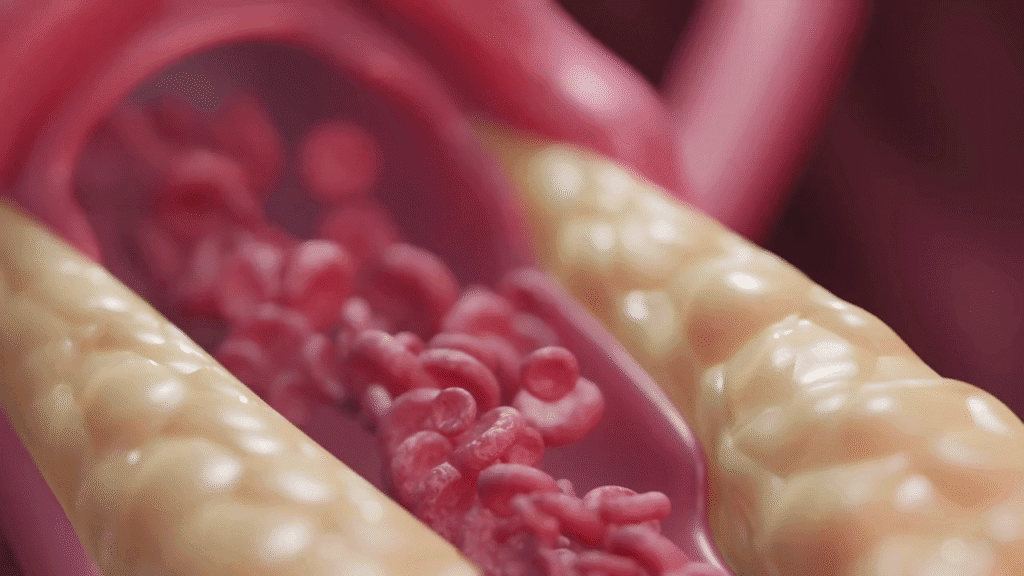
Its main strength is tissue detail. An echocardiogram shows heart structure and movement very well, but MRI can often show what is happening inside the heart muscle itself. That can be useful when looking for scarring after a heart attack, inflammation from myocarditis, inherited cardiomyopathy, or causes of otherwise unexplained heart failure.
When is a cardiac MRI useful?
A cardiac MRI is usually requested to answer a specific clinical question. Common reasons include:
- an echocardiogram has shown something that needs clarification;
- heart failure is present but the cause is uncertain;
- myocarditis or heart muscle inflammation is suspected;
- there is concern about cardiomyopathy, including inherited forms;
- doctors need to assess whether heart muscle is scarred or still viable after reduced blood supply;
- a cardiac mass, congenital abnormality or complex structural problem needs further definition.
The scan is not always the first test. In many patients, the starting point is still a careful history, examination, ECG and echocardiogram. MRI becomes useful when the answer depends on tissue characterisation or accurate measurement of heart volumes and function.
What happens during the scan?
You lie on a table that moves into a large tube-shaped scanner. The scan usually takes around 45-60 minutes. You will hear loud knocking noises, so headphones are used. You may be asked to hold your breath for short periods while images are taken.
Some scans use a contrast agent given through a small cannula in the arm. This helps show scarring or inflammation in the heart muscle. Most people go home immediately afterwards. The scan is not painful, although some patients find the enclosed space uncomfortable.
How the result should be interpreted
A cardiac MRI report is most useful when it is interpreted alongside the clinical history, ECG, blood tests and echocardiogram. The scan may show scar, inflammation or abnormal heart muscle thickness, but those findings still need to be connected to the patient’s symptoms and overall risk.
Dr Mark Cassar is accredited in cardiac MRI, echocardiography and cardiac CT. That can be helpful when deciding which imaging test is most appropriate, and when comparing findings across different tests rather than looking at one scan in isolation.
When to consider private assessment
Private cardiology assessment may be useful if you have been advised to consider cardiac MRI, if previous tests have not explained your symptoms, or if you need a clearer plan after an abnormal scan. Dr Cassar sees patients at Candover Clinic and The Hampshire Clinic in Basingstoke, and The Berkshire Clinic in Reading.
For details of available imaging and consultation options, see Dr Cassar’s cardiology services or the appointments page.