
A detailed scan of the heart and coronary arteries, most often used to look for fatty deposits and narrowing in the arteries that supply the heart muscle.
Cardiac investigation
Cardiac CT and CT coronary angiography
What this test is and when it helps
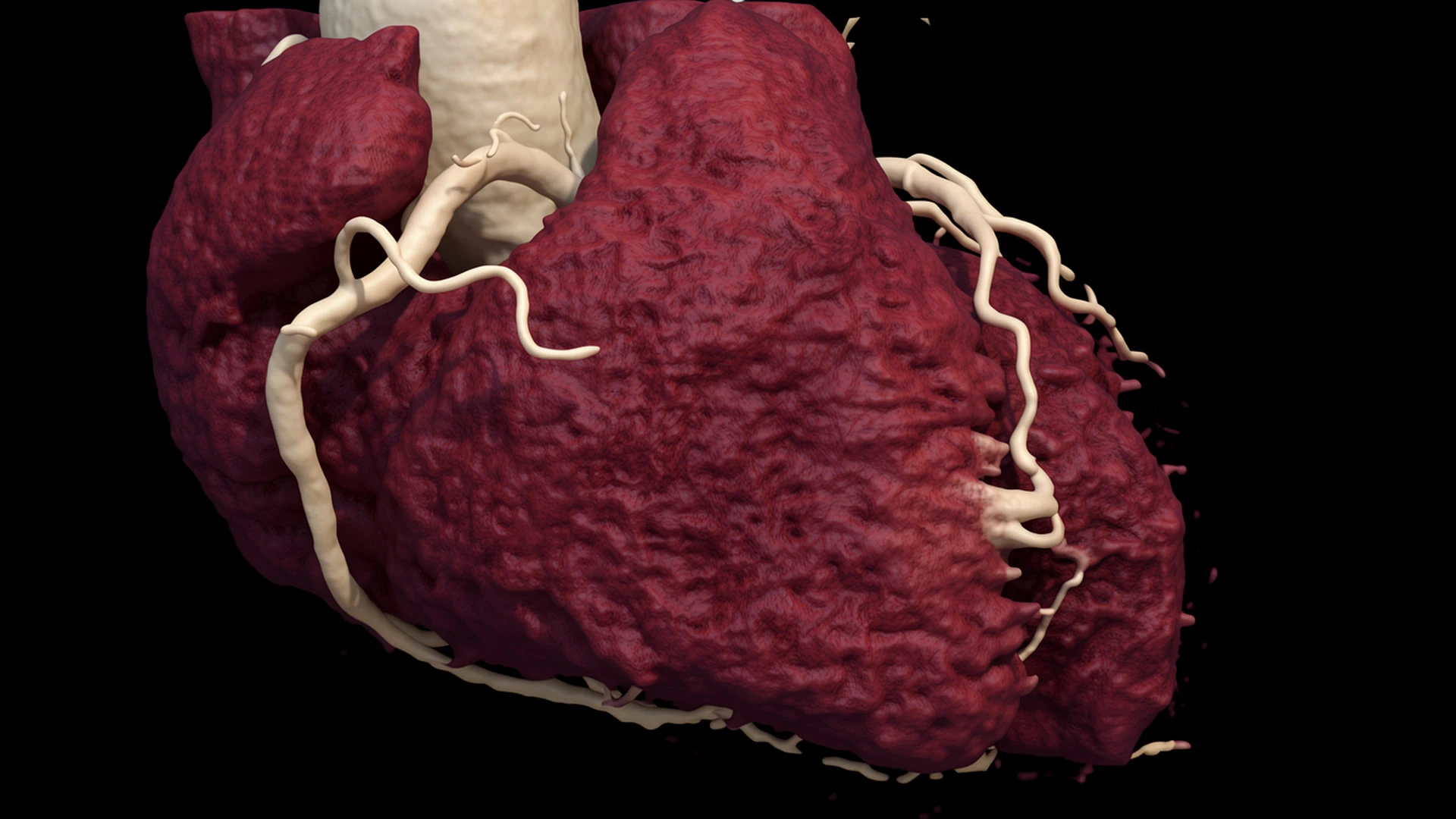
Cardiac CT uses X-rays to take detailed pictures of the heart. The type most often used to investigate chest pain is a CT coronary angiogram, usually shortened to CTCA.
A CTCA shows the coronary arteries, the blood vessels that supply the heart muscle. It can detect fatty deposits, known as plaque, and show whether an artery has become narrowed.
The test may be helpful when chest discomfort or breathlessness could be caused by coronary artery disease. It is also useful when a cardiologist needs to know whether plaque is present, even if it is not causing a severe narrowing.
CTCA is not the right test for every patient. The decision depends on your symptoms, medical history, heart rhythm, kidney function and previous results.
What cardiac CT can show
Coronary plaque
Shows calcified, non-calcified and mixed plaque, including disease that may not yet cause a severe narrowing.
Artery narrowing
Shows where a coronary artery has narrowed and how mild, moderate or severe the narrowing appears.
Cardiac anatomy
Shows the origin and course of the coronary arteries, with selected information about the heart and aorta.
Finding plaque does not necessarily mean that an artery is severely narrowed. Early or mild plaque may still affect how cardiovascular risk is managed, including decisions about cholesterol, blood pressure and smoking.
CTCA and coronary calcium scoring are different tests
A coronary calcium score is a simpler CT scan that does not require contrast. It measures calcified plaque but does not show non-calcified plaque or the inside of the coronary arteries in the same detail as CTCA.
A calcium score of zero can be reassuring in the right setting, but it does not exclude all coronary plaque. This is particularly important in younger people and in patients who have symptoms.
What happens during the scan
You lie on a table that moves through the CT scanner. ECG stickers are placed on your chest so the scanner can take pictures at the correct point in each heartbeat.
A small cannula is placed into a vein, usually in the arm. Iodine-based contrast is injected to make the coronary arteries visible. It is common to notice a brief warm feeling or metallic taste.
You need to lie still and hold your breath for short periods. Clear images are easier to obtain when the heartbeat is reasonably slow and regular. The scanning team may use medication to slow the heart or widen the coronary arteries. Follow the instructions provided for your appointment rather than changing medication yourself.
CT uses a small dose of ionising radiation. Modern scanners adjust the dose to obtain the images needed while keeping exposure as low as reasonably practicable. Tell the team about kidney problems, pregnancy, significant asthma or any previous reaction to contrast.
Understanding the result
The report should explain whether coronary plaque is present, where it is and whether any narrowing appears mild, moderate or severe.
A normal, good-quality CTCA makes important narrowing in the major coronary arteries unlikely. It does not exclude every cause of chest pain, problems affecting very small blood vessels or the possibility of developing coronary disease in future.
A moderate narrowing on CT may or may not reduce blood flow enough to cause symptoms. The result therefore needs to be considered alongside your symptoms, risk factors, examination and other tests. Image quality may be reduced by a fast or irregular rhythm, movement, extensive coronary calcium or some coronary stents.
When another test may be needed
Functional testing
Stress echocardiography or stress cardiac MRI may help when the question is whether a narrowing restricts blood flow.
Invasive angiography
May be needed when CT suggests severe narrowing, images are unclear or treatment may be required during the same procedure.
Other heart scans
Echocardiography is usually better for valves and pumping function. Cardiac MRI is often more useful for heart muscle, inflammation and scar.
How Dr Cassar uses cardiac CT in assessment
Dr Mark Cassar achieved Level 3 accreditation in cardiac CT. He interprets cardiac CT findings alongside the patient’s symptoms, cardiovascular risk and other investigations.
For someone undergoing chest pain assessment, the important question is not simply whether the scan is labelled normal or abnormal. It is whether the amount and pattern of plaque explain the symptoms and whether the result should change treatment.
A moderate narrowing may need another test to assess blood flow. Mild plaque may be more relevant to reducing future risk than to explaining the current symptom. In other cases, normal coronary arteries may allow the assessment to focus on a different cause. The heart scans compared guide explains how CT differs from other tests.
Frequently asked questions
Is cardiac CT the same as CT coronary angiography?
Cardiac CT is the broad term for CT scans of the heart. CT coronary angiography is a particular cardiac CT scan used to show the coronary arteries with contrast.
What can a CT coronary angiogram detect?
It can identify calcified and non-calcified plaque, show where plaque is located and assess whether it appears to narrow a coronary artery.
Is CTCA the same as a coronary calcium score?
No. A calcium score measures calcified plaque without contrast. CTCA uses contrast and shows the inside of the coronary arteries in greater detail.
Does CTCA involve radiation and contrast?
Yes. It uses X-rays and usually requires iodine-based contrast. The team will consider kidney function, pregnancy and any previous contrast reaction before the scan.
Can a normal CTCA rule out coronary artery disease?
A good-quality CTCA showing normal major coronary arteries makes significant narrowing unlikely. It does not exclude disease of very small vessels or every possible cause of chest pain.
Will I need another test afterwards?
Not always. Another test may be helpful if the images are unclear, the scan suggests important narrowing or the result does not fully explain the symptoms.
Discussing whether cardiac CT is appropriate
Dr Mark Cassar can review your symptoms, cardiovascular risk and previous results, then advise whether cardiac CT or a different investigation is likely to answer the clinical question.
Or call 020 3198 9826 or email [email protected]
Not an emergency service. Call 999 for a medical emergency. Do not wait for a private appointment. Medical information disclaimer.