
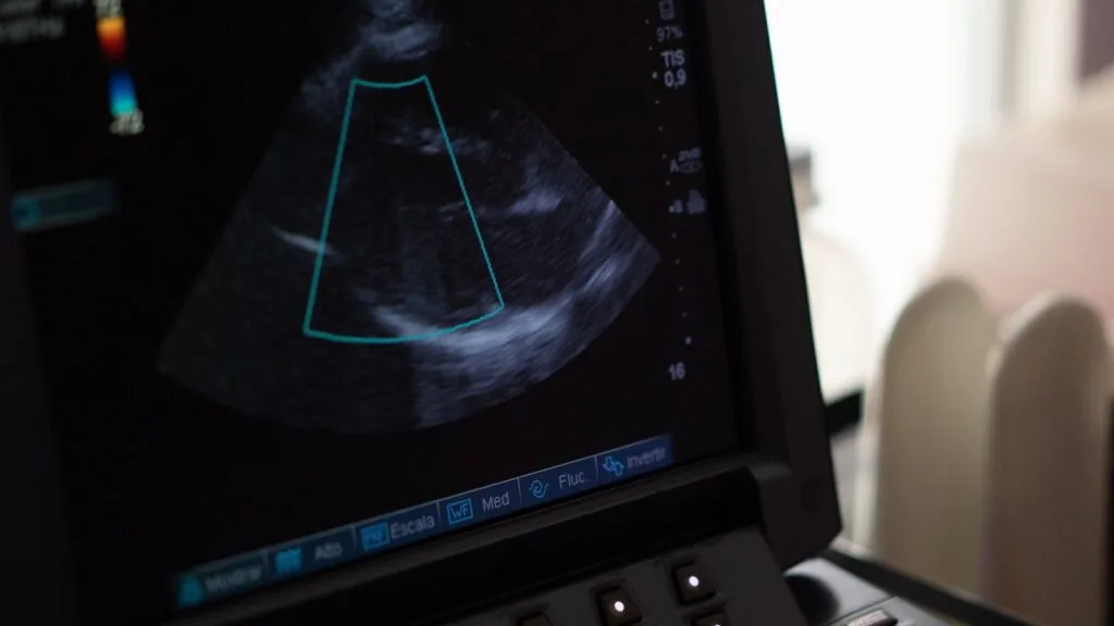
Ejection fraction is the percentage of blood in the left ventricle, the heart’s main pumping chamber, that is expelled with each beat. It is not the percentage of your heart that is working. A healthy ventricle never empties completely, so a figure of 55% does not mean that 45% of the heart has failed.
In current UK heart-failure guidance, an ejection fraction of 40% or less is described as reduced, 41% to 49% as mildly reduced, and 50% or more as preserved. Echo laboratories may use slightly different reference ranges, however. The number therefore needs to be interpreted alongside the rest of the scan, your symptoms and how it was measured.
What does ejection fraction measure?
During each heartbeat, the left ventricle fills with blood and then contracts. Ejection fraction compares the volume ejected with the volume the chamber held when full.
On a transthoracic echocardiogram, this is estimated from ultrasound images of the chamber. On cardiac MRI, the volumes are measured more directly. Both methods are useful, but they do not always produce exactly the same number.
Ejection fraction does not measure the coronary arteries, heart valves, rhythm or how well the ventricle relaxes. Each can be abnormal even when ejection fraction is normal.
What do 35%, 40%, 50% and 55% broadly mean?
UK heart-failure categories provide a useful framework, provided they are treated as ranges rather than absolute verdicts.
- 35% falls clearly within the reduced range. It suggests significantly weakened pumping function and warrants assessment of the cause. It does not, by itself, determine symptoms or outlook.
- 40% is at the upper boundary of the reduced category. A single reading at this level should be interpreted with the scan quality and possible measurement variation in mind.
- 50% meets the guideline definition of preserved ejection fraction, although some echo laboratories use a slightly higher lower limit before describing a result as clearly normal.
- 55% is within the normal range for most laboratories and methods.
An unusually high ejection fraction is not necessarily a sign of an exceptionally healthy heart. A small or thickened ventricle can eject a high proportion of a relatively small volume, so the rest of the scan still matters.
Is there a normal ejection fraction for age?
There is no simple age table that turns a reduced result into a normal one. Hearts become stiffer with age, but clearly reduced pumping function should not be dismissed as normal ageing.
A low ejection fraction is a finding, not the whole diagnosis
A reduced ejection fraction is an imaging finding. Heart failure is a clinical syndrome involving symptoms, such as breathlessness, fatigue or ankle swelling, together with objective evidence that the heart is responsible.
The two often occur together, but reduced left ventricular function can be found before symptoms develop. That still deserves assessment, but it is not identical to symptomatic heart failure.
The first question is whether the number is reliable and whether it fits the symptoms, the ECG and the rest of the scan. A surprising result in someone who feels well and had technically difficult images is interpreted differently from the same number in someone who is breathless with a visibly enlarged ventricle.
Can heart failure occur with a normal ejection fraction?
Yes. In heart failure with preserved ejection fraction, often abbreviated to HFpEF, the ejection fraction is preserved but the ventricle may be stiff and fill at raised pressures.
Diagnosing HFpEF requires symptoms plus objective evidence of abnormal structure or filling. A normal ejection fraction alone neither confirms nor excludes it. This is an important source of false reassurance when breathlessness is attributed to age or fitness because the report says that pumping function is preserved. A structured breathlessness assessment looks beyond the single number.
Why can two scans give different results?
Echocardiography depends on image quality and on how the borders of the ventricle are traced. Two experienced readers can produce slightly different estimates from the same study.
Heart rhythm also matters. In atrial fibrillation, the amount of blood ejected varies from beat to beat. Blood pressure, hydration and how the heart is loaded on the day can also influence the result.
A difference of only a few percentage points may reflect measurement variation. Before concluding that the heart has improved or deteriorated, compare the technique, rhythm, loading conditions and image quality. Trends are most useful with the same imaging method and a comparable technique.
When does cardiac MRI help?
Cardiac MRI measures ventricular volumes without relying on the same geometric assumptions as echocardiography. It can also characterise the heart muscle, helping to look for scar from a previous heart attack, inflammation or certain heart-muscle conditions.
It can be particularly useful when echo images are poor, when a borderline measurement could change management, or when the cause of a weakened ventricle is unclear. A small difference between an echo result and an MRI result does not necessarily mean that the heart changed between tests. The methods measure the ventricle differently.
What commonly causes a low ejection fraction?
The clinically useful task is to identify the cause, not simply relabel the percentage. Possible causes include:
- coronary artery disease or a previous heart attack
- cardiomyopathy or myocarditis
- a sustained fast heart rhythm
- significant heart valve disease
- longstanding high blood pressure
- alcohol and certain other toxins
- some cancer treatments
- inherited heart-muscle conditions
- thyroid, endocrine or metabolic problems
Which possibilities need investigation, and in what order, depends on the history, ECG and other scan findings.
Can ejection fraction improve?
It can. When the cause is reversible or responds to treatment, ejection fraction may rise. Examples include controlling a persistently fast rhythm, stopping excess alcohol, or treating heart-muscle weakness.
Improvement is not guaranteed, and treatment depends on the cause and the wider clinical picture. This is why establishing the reason for the reduced function is more useful than focusing on the percentage alone.
Can ejection fraction predict life expectancy?
Not by itself. Prognosis also depends on symptoms, the underlying cause, right-heart function, rhythm, kidney function, other illnesses, response to treatment and the direction of change over time.
Two people with the same ejection fraction can have very different clinical courses.
What usually happens after a reduced result?
Assessment commonly includes a careful history and examination, an ECG and relevant blood tests. Further imaging, coronary assessment or rhythm monitoring may be considered when these would help establish the cause.
If the finding was unexpected, it is reasonable to ask about image quality and whether confirmation is needed. The next test should answer a specific clinical question.
When should you seek urgent help?
Call 999 or attend A&E for severe or sudden breathlessness, persistent heavy chest pain, collapse, or feeling acutely very unwell. For concerning worsening symptoms that are not immediately life-threatening, such as increasing breathlessness or new swelling, contact NHS 111 or your GP for urgent advice rather than waiting for a routine appointment.
Reviewing an ejection-fraction result
Dr Mark Cassar can assess concerns about an unexpected or reduced ejection fraction in clinic, review previous reports and images, and arrange further tests where appropriate. If you would like a personalised review, the appointments page has details on arranging a consultation.
Frequently asked questions
What is a dangerously low ejection fraction?
There is no single dangerous cut-off. Values of 40% or below are classed as reduced, and lower values generally indicate more marked pumping impairment. Risk also depends on the cause, symptoms, rhythm and response to treatment, so a low result needs clinical interpretation rather than an isolated label.
Can ejection fraction improve?
Yes, in many cases. Improvement is most likely when the cause is reversible or responds to treatment. Repeat scans should use a comparable method where possible so that genuine change can be distinguished from measurement variation.
Can you have heart failure with a normal ejection fraction?
Yes. In heart failure with preserved ejection fraction, the ejection fraction is preserved but the ventricle may be stiff and fill at raised pressures. Diagnosis requires symptoms plus objective evidence of this problem.
Is 55% a normal ejection fraction?
For most laboratories and methods, yes. A result of 55% is within the normal range, but it should still be read alongside the rest of the report, including the valves, chamber sizes, wall thickness and right-heart findings.
Sources and further reading
- NICE: chronic heart failure recommendations, updated September 2025
- British Heart Foundation: heart failure
- British Heart Foundation: understanding an echocardiogram report
- British Society of Echocardiography: normal reference intervals for adult echocardiography
- NHS England: managing heart failure at home