
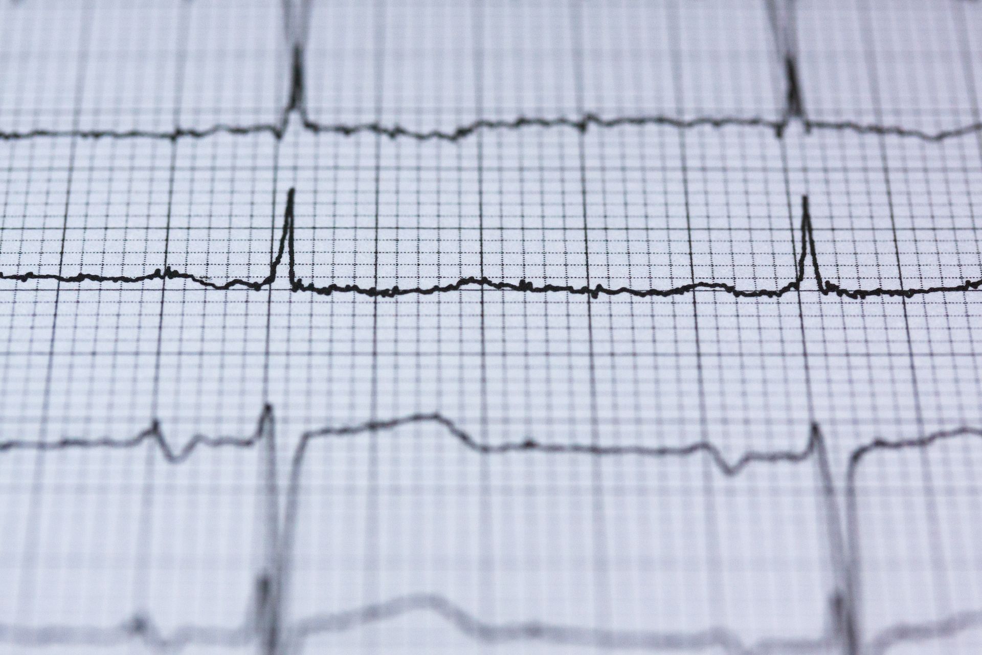
ECG series, part 1 of 3. Part 2: left bundle branch block. Part 3: right bundle branch block.
Being told your ECG is "abnormal" is worrying, but it is a broad label. It covers findings that are harmless variations of normal as well as findings that need further tests. Many ECG machines also print an automatic interpretation that can flag minor variations unnecessarily. The useful questions are what exactly the ECG shows, whether it is new compared with any earlier tracing, and whether it fits your symptoms.
What an ECG records
An ECG records the heart's electrical activity for about ten seconds, using stickers on the chest, arms and legs. It shows the heart rate and rhythm, how the electrical signal spreads through the heart, and indirect clues about the heart muscle. It does not show the heart arteries or valves directly, and it records only the ten seconds it is running.
What a normal ECG looks like, and what makes one abnormal
Each heartbeat produces a repeating pattern of waves. The small P wave shows the upper chambers activating. The tall, narrow QRS complex shows the main pumping chambers activating. The T wave shows the heart muscle resetting before the next beat. A standard ECG views this pattern from 12 directions, called leads.
An ECG is called abnormal when the rate, rhythm, timing or shape of these waves falls outside the expected range. Some of those differences are normal for a particular person, for example because of fitness, age or body shape. Others point to a condition that needs looking at.
"Sinus rhythm, abnormal ECG"
Many reports say "sinus rhythm" alongside "abnormal ECG". Sinus rhythm means the heartbeat is starting from its normal place, which is reassuring. The "abnormal" part refers to something else on the tracing, such as the shape of a wave, and needs reading in its own right.
Why the machine's printed report needs checking
The printed interpretation is produced by software, applying fixed measurement rules to the tracing. Automatic reports can flag a normal variation as abnormal, and can occasionally miss something a doctor would notice. Phrases such as "possible", "consider" or "borderline" are common and often turn out to be normal variations once a clinician reads the tracing itself. The tracing, not the printed summary, is what matters, which is why a doctor's review of an abnormal result is worthwhile even when the printed wording sounds alarming.
Common findings on an abnormal ECG
- A slow or fast heart rate. Often normal, for example a slow rate in a fit person or a fast rate when anxious.
- Extra beats (ectopics). Very common and usually harmless; see the ectopic beats article.
- Bundle branch block. A delay in the electrical pathway on the left or right side of the heart; see parts 2 and 3 of this series.
- Signs of a thickened heart muscle. Larger ECG waves can suggest left ventricular hypertrophy, but the ECG often over- or under-estimates it, so an echocardiogram is used to check.
- Signs of a previous heart attack. Some patterns suggest an old area of heart muscle damage, which may not have been noticed at the time.
- T wave inversion. Explained below.
- Atrial fibrillation. An irregular rhythm that needs its own assessment; see the atrial fibrillation page.
What does T wave inversion mean?
The T wave is the part of each heartbeat that shows the heart muscle resetting. Normally it points upwards in most leads. T wave inversion means it points downwards.
In some leads it is a normal finding. An inverted T wave is expected in lead aVR, is common in V1, and is often seen in lead III on its own, so these particular patterns are usually not a cause for concern. T wave inversion across V1 to V3 can also occur as a normal variant in children and in some trained athletes, but its significance in an adult depends on exactly which leads are affected, how deep the inversion is, the rest of the ECG, your age, your symptoms and any earlier tracings for comparison. Inversion extending beyond V1, or occurring alongside other ECG changes, should not be dismissed simply because the person is young or female.
In other leads, or when it is deep or new, T wave inversion can point to reduced blood supply to the heart, a previous heart attack, strain from high blood pressure or a valve problem, or a heart muscle condition such as cardiomyopathy. Which leads are affected, your age and symptoms, and comparison with earlier ECGs decide which applies. New T wave inversion with chest pain needs urgent assessment.
Can an abnormal ECG mean a heart attack?
An ECG can show signs of a heart attack that is happening now, or of one in the past. But an abnormal ECG does not usually mean you have had a heart attack, and a normal ECG does not rule one out, which is why hospitals combine the ECG with blood tests. If you have chest pain that does not go away, or chest pain with sweating, sickness or breathlessness, call 999 rather than waiting for an ECG result.
When to seek urgent help
Call 999 for symptoms of a possible heart attack, such as sudden chest pain or discomfort that does not go away, pain spreading to the arm, jaw or back, or chest pain with sweating, sickness or breathlessness. Call 999 for severe breathing difficulty.
If someone faints, call 999 if they do not wake within a minute, have not fully recovered, have chest pain or palpitations, are seriously injured, have a seizure, or faint during exercise or while lying down. Do not drive yourself to A&E.
Even if you have recovered, a blackout alongside an ECG abnormality such as a conduction problem needs urgent medical assessment. Contact your GP or NHS 111 the same day and mention the ECG finding rather than waiting for a routine appointment. If you are awaiting specialist cardiovascular assessment after a blackout, do not drive until you have been advised it is safe. If you have otherwise had a faint and recovered fully, arrange a GP assessment.
Many abnormal ECG findings can be reviewed at a planned appointment, but some need urgent assessment even without symptoms. Follow any urgent advice given by the clinician who arranged or reviewed your ECG; do not use the absence of symptoms as reassurance on its own.
What usually happens next
The first step is to look at the tracing itself and compare it with any earlier ECGs. A finding that has been present unchanged for years means something different from one that has newly appeared, though a newly discovered finding may simply mean no earlier ECG exists for comparison rather than that it has just developed. Depending on what is found, the next test may be:
- an echocardiogram to check the heart's structure and pumping
- a 24-hour ECG or longer heart monitor if the question is about rhythm, with the duration tailored to how often any symptoms occur
- cardiac CT or stress imaging if the question is about the heart arteries
- blood tests, such as thyroid function or salts
Many people need no further tests once the ECG has been reviewed.
See Dr Cassar about this
Dr Mark Cassar can review your ECG alongside your symptoms and previous results, and advise whether further tests would help.
- Adults (18 and over), at Candover Clinic and The Hampshire Clinic, Basingstoke
- First appointments usually within a week
- Initial consultation £250, follow-up £150; tests are charged separately
- Call 020 3198 9826 or email [email protected]
Frequently asked questions
What does an abnormal ECG mean?
That something on the tracing falls outside the expected range. It ranges from harmless variations to findings that need further tests, so it needs reading by a doctor.
Is an abnormal ECG dangerous?
Usually not on its own. It is a clue rather than a diagnosis, and its importance depends on what it shows and on your symptoms.
Can an ECG be wrong?
Both the recording and its interpretation can be affected by errors. Movement, poor electrode contact or incorrect electrode positioning can distort the tracing, and the automatic interpretation printed on it can over-call minor findings. A clinician checks the ECG itself, and may repeat it if the recording is unclear.
What causes an abnormal ECG result?
Common causes include fitness, age, extra beats, high blood pressure, a delay in the heart's wiring, a previous heart attack, and heart muscle or valve conditions. Electrode position and movement can also distort a tracing.
Can an abnormal ECG become normal?
Yes. Some findings change with heart rate, posture, salts in the blood or treatment, and some apparent abnormalities disappear when the ECG is repeated with careful lead placement.
Can anxiety cause an abnormal ECG?
Anxiety can speed up the heart rate and occasionally cause minor changes, but it should not be assumed to explain an abnormal ECG without a review.
Does an abnormal ECG mean heart disease?
No. Many abnormal findings are variations of normal. Some need an echo or monitor to decide.