
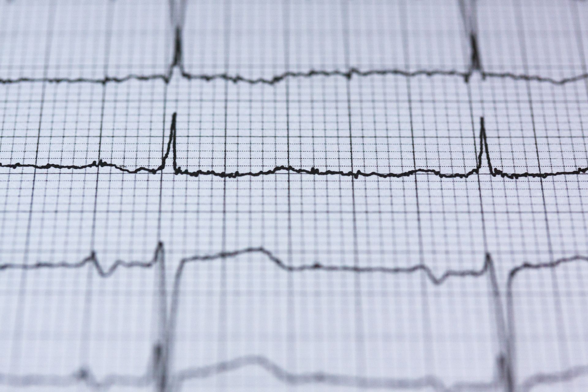
ECG series, part 2 of 3. Part 1: abnormal ECG result. Part 3: right bundle branch block.
Left bundle branch block, or LBBB, means the electrical signal reaches the left side of the heart more slowly than usual, so the main pumping chamber is activated late. You cannot feel it, and many people are told about it only after a routine ECG. Unlike right bundle branch block, LBBB is more often linked to an underlying heart condition, so finding it usually leads to a heart scan even if you feel well. In many people that scan is normal.
What LBBB is
Each heartbeat starts with an electrical signal at the top of the heart. The signal travels down two pathways, the right and left bundle branches, so that both pumping chambers contract together. The left bundle branch carries the signal to the left ventricle, the heart's main pumping chamber.
In LBBB, that pathway conducts slowly or not at all. The signal still reaches the left ventricle, but by a slower route through the heart muscle. The two sides of the heart then contract slightly out of step, which produces a wide, characteristic pattern on the ECG.
Complete, incomplete and intermittent LBBB
An ECG report may describe LBBB in different ways:
- Complete LBBB is the full pattern, and the one usually meant by "LBBB".
- Incomplete LBBB shows a similar shape with a smaller delay. It is often assessed in the same way, because the same conditions can cause it.
- Intermittent or rate-related LBBB comes and goes, often appearing only when the heart rate rises. It may be seen on one ECG and not the next, or during a heart monitor recording or exercise.
Right and left bundle branch block compared
Incomplete right bundle branch block (RBBB) is common in younger and fitter people, and is usually harmless when the rest of the heart is normal. LBBB is less common, becomes more frequent with age, and is more often linked to high blood pressure, heart muscle disease or coronary artery disease. That difference is why isolated incomplete RBBB often needs no further tests, while LBBB is usually followed by a heart scan. The right bundle branch block article explains RBBB in more detail.
What can cause it?
- Long-standing high blood pressure, which thickens and strains the heart muscle; see high blood pressure.
- Coronary artery disease, including damage from a previous heart attack; see coronary artery disease.
- Heart muscle conditions (cardiomyopathy), where the left ventricle is enlarged, weakened or thickened.
- Aortic valve narrowing; see aortic stenosis.
- Inflammation of the heart muscle (myocarditis), rarely, sometimes after a viral infection.
- Age-related wear of the conduction system, often with no other heart problem.
- Heart procedures, such as aortic valve replacement, which can affect the nearby conduction pathways.
Sometimes no cause is found and the heart is otherwise normal.
Symptoms
LBBB itself does not usually cause symptoms. When symptoms are present, they normally come from the underlying condition: breathlessness, tiredness or ankle swelling if the heart muscle is weak; chest tightness on exertion if the heart arteries are narrowed; or dizziness and fainting if the heart's wider electrical system is affected and the heart rate drops.
It helps to mention any of these at your assessment, even if they seem minor, because they guide which tests are useful.
When to seek urgent help
Call 999 for sudden chest pain or discomfort that does not go away, particularly with sweating, nausea or breathlessness, or for severe difficulty breathing. A newly found LBBB with chest pain needs emergency assessment, since LBBB can make a heart attack harder to recognise on an ECG.
If someone faints, call 999 if they do not wake within a minute, have not fully recovered, have chest pain or a pounding or irregular heartbeat, are seriously injured, have had a seizure, or faint during exercise or while lying down.
If you have a blackout and recover fully, but LBBB or another ECG abnormality has been found, this still needs urgent medical assessment the same day, rather than waiting for a routine appointment; mention the ECG finding when you contact your GP or NHS 111. This is usually arranged as an urgent specialist cardiology referral. Until that assessment has taken place, do not drive.
Why a heart scan is usually recommended
An echocardiogram is usually the first test. It shows whether the heart muscle, valves and pumping function are normal. If the echo is unclear, or if the heart muscle is weak and the cause is uncertain, cardiac MRI can show scar, inflammation or other heart muscle conditions.
LBBB also changes how other tests are read. The ECG changes it causes make an exercise ECG unsuitable on its own for diagnosing reduced blood supply to the heart, although it may still be useful for other purposes. If heart artery disease needs checking, the cardiologist will choose cardiac CT or an appropriate stress imaging test; the imaging method and stress protocol matter, because LBBB can also affect some imaging results. If dizziness or fainting is part of the picture, a 24-hour ECG or longer heart monitor looks for intermittent slowing of the heart rhythm. The length of monitoring is tailored to how often symptoms occur; infrequent episodes may need a longer recording or other specialist testing, since a normal short monitor does not rule out an occasional slow rhythm.
New or long-standing LBBB
It makes a difference whether LBBB is new compared with an earlier tracing. If an earlier ECG was normal, a new LBBB suggests something has changed and deserves prompt assessment. If it has been present for years on old tracings and the heart was normal when last checked, the question is usually whether anything has changed since. If there is no earlier ECG to compare with, "newly found" is not the same as "newly developed": the block may have been present for some time without being picked up. Bring any previous ECGs or letters to your appointment if you can.
Does LBBB need treatment?
There is usually no treatment specifically needed for isolated LBBB; treatment depends on the underlying condition, such as controlling blood pressure, treating narrowed arteries or treating a heart muscle condition.
In some people with heart failure, a weak left ventricle and LBBB, a specialist may recommend a particular type of pacemaker that resynchronises the two sides of the heart, called cardiac resynchronisation therapy, once heart failure has been assessed and appropriately treated. In a smaller number of people, the out-of-step contraction caused by LBBB can itself weaken the heart over time, which is another reason the heart's pumping is rechecked periodically.
A conventional pacemaker is not usually needed for isolated LBBB. One may be needed if more serious conduction disease is found, such as intermittent or advanced heart block. Some patterns of conduction disease warrant pacing even without symptoms. Resynchronisation pacing for selected people with heart failure is a separate decision, as described above.
Outlook
The outlook depends on the underlying cause, how well the heart pumps and the wider conduction system. Many people with LBBB and an otherwise normal heart scan do well. Follow-up is tailored to the initial findings and any new symptoms, rather than one fixed schedule for everyone. When LBBB occurs alongside a weak heart muscle, the outlook depends mainly on that condition and how it responds to treatment.
See Dr Cassar about this
Dr Mark Cassar can review your ECG and any previous heart scans, assess possible causes of left bundle branch block, and advise on further tests or follow-up.
- Adults (18 and over), at Candover Clinic and The Hampshire Clinic, Basingstoke
- First appointments usually within a week
- Initial consultation £250, follow-up £150; tests are charged separately
- Call 020 3198 9826 or email [email protected]
Frequently asked questions
Is left bundle branch block serious?
It can be harmless, but it is more often linked to heart disease than RBBB, so it should be checked with a heart scan. If the scan is normal, the outlook is usually good.
Can you die from left bundle branch block?
LBBB itself is not usually dangerous. The risk comes from any underlying condition, such as a weak heart muscle or narrowed arteries, which is why finding the cause matters.
Can left bundle branch block cause a heart attack?
No. LBBB does not cause heart attacks, but it can be a sign of coronary artery disease, and it makes a heart attack harder to see on an ECG.
Can left bundle branch block go away?
Intermittent or rate-related LBBB comes and goes. Persistent LBBB usually remains, although it occasionally resolves when a reversible cause is treated.
Can a virus cause left bundle branch block?
Rarely. Inflammation of the heart muscle after a viral infection can affect the conduction system. Cardiac MRI can help show whether this has happened.
Does caffeine affect left bundle branch block?
Caffeine does not cause LBBB, and there is no need to avoid moderate amounts because of LBBB alone.
Is there a diet for left bundle branch block?
There is no specific diet for LBBB. A heart-healthy diet helps with causes such as high blood pressure and coronary artery disease; see the Mediterranean diet and heart health.
Can I exercise with left bundle branch block?
If your heart is otherwise normal and you have no symptoms, LBBB is not usually a reason to stop exercising. New breathlessness, chest pain or dizziness on exertion should be assessed before you continue.
Can I drive with LBBB?
For car and motorcycle drivers, LBBB alone does not require DVLA notification if there is no other disqualifying condition. Bus, coach and lorry drivers must tell DVLA if they have LBBB. Blackouts or other heart conditions may mean you need to stop driving and meet separate requirements; your doctor will advise what applies to you.
Will I need a pacemaker?
Not usually. A pacemaker may be considered if the heart's electrical system becomes more widely affected, which does not always cause a slow heart rate, or in selected people with heart failure, to resynchronise the heart.